


Five reasons to adopt microscopy to help make better agronomic decisions
When we use biologicals in farming or gardening, it’s critical to use a microscope to check on those microbes. You could easily just be spinning your wheels, thinking you are doing something of value, but meanwhile we are adding no value; or, worse, you’re doing harm.
Consider a farmer using Korean Natural Farming (KNF) techniques. KNF has a great success record in many contexts, but it’s not infallible. It could be that a high water table is adding contamination to the system. It could be contamination in the well water. Eutrophication from runoff fertilizers combined with glyphosate residues and contaminated manure has created an ecosystemic catastrophe in many areas. There could be glyphosate and other contaminants in the animal feed that is turned into compost. And it could always be user error in the composting process.
The point is that if we don’t look, we don’t see. If we don’t test, we don’t know what, or if, anything is wrong. We need to look at our final product, but also the ingredients going into them. If we can only react when a macro-expression of the problem presents itself, we are just waiting for something bad to happen: a field speckled with blight, discoloration of leaves, chlorosis, insect damage, etc.
These are symptoms of biological or mineral chinks in the armor — but we could have seen these issues early on, as they developed, before they turned into problems we can see with the naked eye. We could have also foreseen problems if we were using a plant sap analysis meter and saw the dip in the plant’s key minerals.
Here are the top five reasons soil microscopy matters.
#1 You Can Catch Problems Early
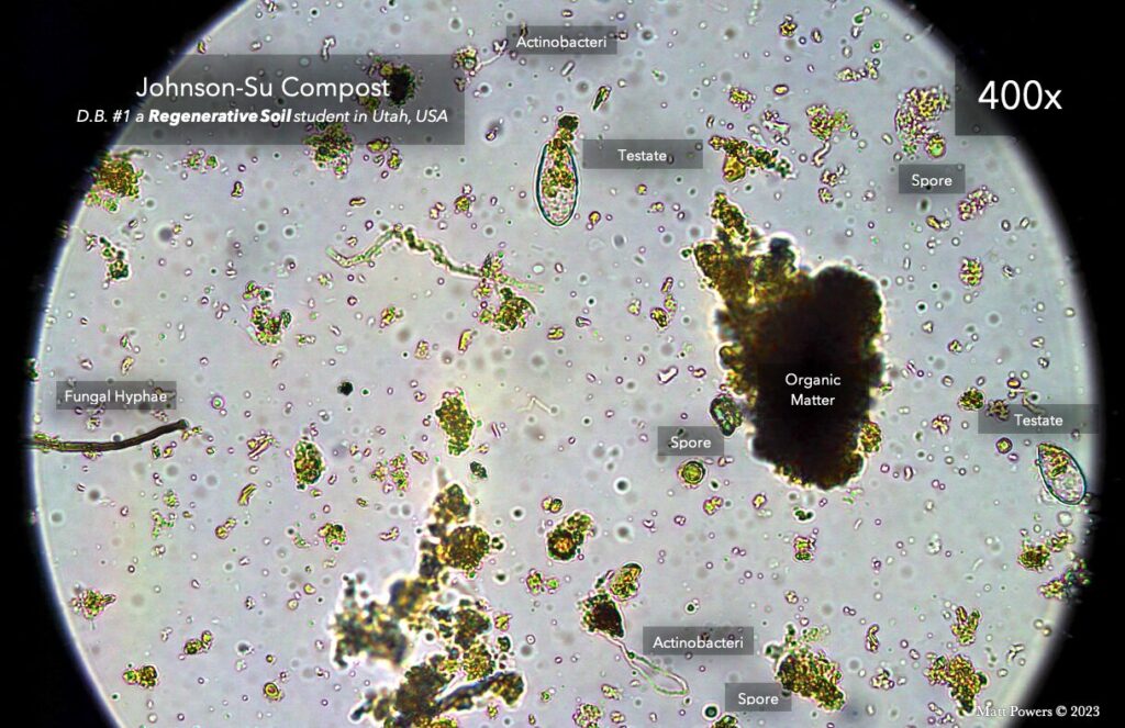
With a microscope, you can see if the compost is anaerobic, empty of life, or imbalanced or incomplete biologically. You can also see pathogens before they are visible to the naked eye. Fungal spores can be identified or at least narrowed down to a handful of lookalikes with keys and global spore atlases, so you can look things up and see what you likely have and react accordingly. Fusarium and alternaria spores are easy to spot, but for some things you’ll want to see more of the life cycle. The stage prior to release of the spore itself can sometimes be the best time to ID some fungi like aspergillus species, which have some beneficial but mostly pathogenic members.
You can also ID the first symptoms you see and create a tailored response, but this is less effective — an ounce of prevention is worth a pound of cure. We can vet our compost on its way in; we can also check our early spring soils. If a plant is slowing down its production of root hairs, we know the plant is either running low on photosynthetic sugars or on microbes to feed into rhizophagy. It could be the soil is dead in this new area, that there is a pocket of biocide residue, or that the plant is not producing enough sugars due to a limiting factor preventing full photosynthesis. This could be a mineral deficiency or simply a lack of light (it could be late season). That’s why we look at multiple plants from multiple areas to see what the trends are across samplings.
Without the right biology in place, a simple seasonal lack of sugars can trigger the expression of late-season blights like fusarium and alternaria since they can hide in plants as endophytes until this specific time and opportunity to turn pathogenic. After that they turn saprophytic, digesting the plant material, forming spores, and waiting for next season to start all over again. This is why it’s worth it to just look.
#2 Most Things Are Easy to See
It takes time and practice, but once you are acquainted with the space, you can easily assess most soil, compost, roots, and more, with some important caveats. I teach my students to be as non-definitive as possible while doing the microscopy part — to leave options of interpretation open until they complete other tests.
There are bacteria that form hyphae that look like fungi, but their hyphae break up into cocci and bacilli shapes. The pathogenic E.coli looks identical to non-pathogenic E.coli, and there can be a large number of bacteria too small to be seen with light microscopy. We have to take a humble stance in this space, avoid using manual quantification as a leading indicator, and recognize we are looking at samplings of a vast space with a diversity that is continuously iterating itself: nature is always adapting and changing.
Mycorrhizal fungi is always changing its genes in response to its environment, making it “new” constantly. This is the ultimate fallacy of GMO thinking — it wants to hold life still, but that’s not how life works. It’s important to lead with humility and observation in tandem with other tests.
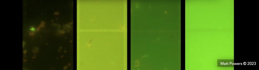
DNA testing has a similar problem — it’s a snapshot of a moving picture and will change with time and environmental conditions. This is why I’ve adapted new testing methods like the biological viability test (to see the relative ratio of living to dead bacteria). We can use the dye as a side-by-side comparison between different samples to see which is more or less biologically rich. It’s much easier now to see which sample has more life in it (the brighter it is, the less life.)
The reality is that most things are easy to identify to their morphological group: bacteria, fungi, protozoa, nematodes, microarthropods, diatoms, algae, etc. Nematodes are categorically separated by their mouthparts, though there is evidence that they can adapt their mouthparts in presence of different food sources, or because of an abundance of males (they turn into predators and grow teeth), or for no reason at all that we can yet tell. Adaptation is the rule here: adapt or die, and the most adaptable are the most abundant and dominant globally (and nematodes are the most abundant animal on earth).
This being said, many folks have not been trained for more advanced applications like identifying mycorrhizal fungi, fungal pathogens, and rhizophagy, and some focus on quantification using multiplication, which leads to large swings of divergence and error This is why many folks have switched to ratios.
#3 Solving the Mycorrhizal Fungi Inoculant Predicament
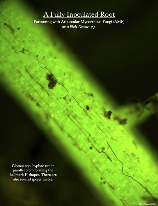
When we can see, we can verify: did you know that everyone has been using mycorrhizal fungi as an inoculant and never even verifying if it was successful for years now? And did you know that these fungi are regenerative in nature? That means that mycorrhizal fungi come back every single year on their own. We just have to encourage them back by avoiding phosphate fertilizer, which inhibits them, and avoiding nitrogen fertilizer, which burns out plant roots (which are the inoculant for the next season).
Because we can now see fungi and their effects, we can compare inoculations, verify inoculants are actually working, and even evaluate inoculants before use or purchase. My vision for the future is to help farmers everywhere get mycorrhizal fungi back in their soils and for them to be fully inoculated every season without any added inputs.
#4 You Can Do It at Home
It’s DIY and easier than you think. If you have some desk space that can get a little dirty and be cleaned up easily, you can do this. It takes a bit of learning, but within a few months of training and practice, you will be comfortable with the microscope.
If you are using eyepieces, though, you may never feel comfortable — I never recommend these because of the damage they do to the eye. That means I always recommend a high-quality camera and monitor to view the microscope images. Using a camera, you can also take pictures or record video of what you are seeing and share it with anyone anywhere.
#5 It’s Fast
You can get answers fast if you know what to look for and how to operate the microscope. If it’s dead, you can find out in only a few minutes — no need to pay for any outside tests. If it’s amazing compost or soil, it’s also just as readily apparent.
It’s also fast to learn how to do it. I teach folks in a 20-week course, but the live lab sessions are only for the second half, so that’s just 10 weeks of intensive training.
What Microscopy Can’t Do

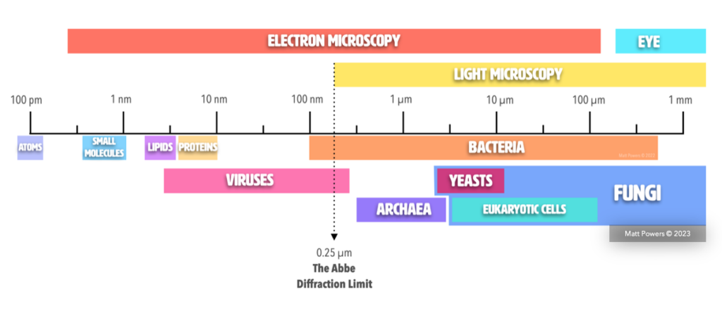
Microscopy has limitations. Light microscopes cannot see almost all viruses (there are actually giant viruses, “giruses,” that can be seen!). It can’t differentiate between pathogenic and nonpathogenic E.coli. It cannot see atoms or molecules or electrons, etc. It can only see a limited bandwidth — electron microscopy goes much further but has its own limitations.
Light microscopy cannot even see all the bacteria. 1000x magnification is the limit of resolution for light microscopy. Epifluorescence can theoretically go slightly higher, but that’s the limit. This is why counting bacteria manually is silly — we cannot see them all; this is why the biological stain is helpful.
Together Is Always Best
Other rests are vital to understanding our microscope results. They can help build certainty, debunk, or discover the root cause of things. Remember: pH determines the form of nitrogenous waste released by microbes, but microbes have the greatest effect on pH and Eh, so we can evaluate the effects of our microbes quite often using simple tests like pH and nitrate tests. But it goes beyond that now: because of the new Croptix meter and Nutriscope meters, we now have ways of verifying our work at the plant-sap and nutrient-density levels, to combine with yield data. Senseen also shares Eh, pH, and Brix, and they are working on a liquid and dry material version of their meter.
These meters change the game entirely — we now have full-cycle testing from the soil to the compost to roots to sap to the fruit, and we can convey that to our customers or buyers, or use it to verify that we are feeding our family the best. But these tests only see nutrients, minerals, and levels like pH/Eh — they cannot see the biology nor how living or dead the soil is, nor what is missing from the biology. These are relatively easy to assess with a microscope and then treat.
The future is to showcase the entire picture: total digestion soil tests, soluble mineral soil tests like M3 or Albrecht, clay/sand/silt/SOM, slake testing, Microbiometer (F:B ratio), plant sap analysis, plus yield numbers — alongside microscopy, sap, roots, and inputs for the crop. In this way, we will be able to witness collectively what works best for a given crop in a given climate in a given soil type. This is my vision with the R-Soil database. Each aspect matters — each relates to other core aspects in fundamental ways. We are viewing a holistic reality, so we have to think and test holistically to even attempt to grasp it.
There’s a reason doctors, researchers, and scientists all over the world are using microscopes in their daily work: there’s nothing else like it that does the job better. There may be new cameras, new monitors and screens, better optics, and new lighting methods, but we are still just magnifying: making small things larger. That alone is a very powerful and useful ability that has stood the test of time and will continue to do so. We’re just finally applying it to gardening and agriculture to make better decisions.